File:Showcase.jpg
Examples of various reconstructed multi-view datasets
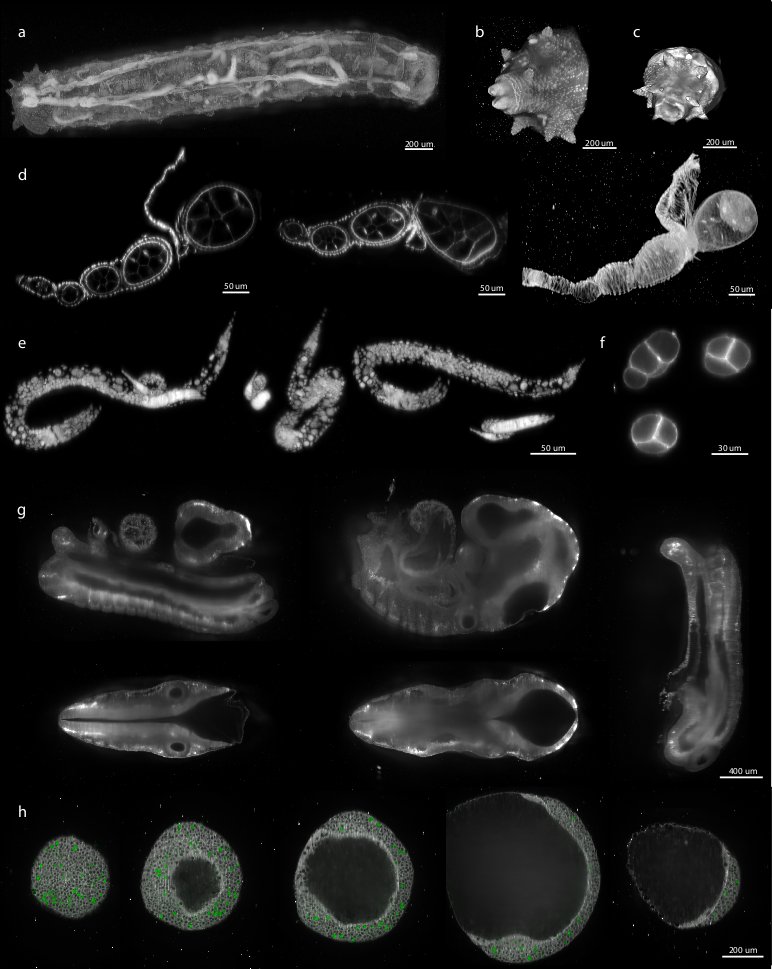
(a) Maximum intensity projection of wild-type Drosophila third instar larva imaged from 22 views with 10× objective. 3d rendering of the posteriors tips of the Drosophila third instar (b) and second instar (c) larvae imaged from 8 views with 10× objective. (a-c) The signal comes from autofluorescence of the larval cuticle. (d) Two perpendicular sections and maximum intensity projection of Drosophila ovariole stained with rhodamine-phalloidin (Molecular Probes) to visualize membranes and imaged from 12 views with 40× objective. (e) L3 and L1 larval C. elegans worm stained with DNA dye; imaged and reconstructed from 8 SPIM views acquired with 40× objective. (f) Sections through single time-point of 6-view, time-lapse recording (40× objective) of C. elegans expressing PH-domain-GFP fusion marking the membranes of 4 cell stage embryo. The C. elegans images highlight the isotropic resolution of the reconstructed volumes. (g) Sections through 9.5 days old fixed, wild-type mouse embryo recording autofluorescence from 19 views with 10× objective. (h) Five planes through fixed zebra fish embryo labeled with membrane marker and cell division marker imaged from 10 views with 20× objective.
File history
Click on a date/time to view the file as it appeared at that time.
| Date/Time | Thumbnail | Dimensions | User | Comment | |
|---|---|---|---|---|---|
| current | 11:44, 27 May 2010 | | 772 × 969 (96 KB) | Axtimwalde (talk | contribs) | Examples of various reconstructed multi-view datasets (a) Maximum intensity projection of wild-type Drosophila third instar larva imaged from 22 views with 10× objective. 3d rendering of the posteriors tips of the Drosophila third instar (b) and second |
- You cannot overwrite this file.
File usage
The following page links to this file: